Clinical Tests of Tibialis Posterior Tendinopathy

A recent study aimed to explore the relationship between ultrasound imaging findings and clinical tests for tibialis posterior tendinopathy.
We reviewed this study in the latest issue of our Research Reviews – where industry experts break down the most recent and clinically relevant studies, for immediate application in the clinic.
What you’ll read below is a snippet from the review.
Like the sound of these Research Reviews? – Learn more HERE
Back to the study!
STUDY TITLE: Clinical tests of tibialis posterior tendinopathy: are they reliable, and how well are they reflected in structural changes on imaging? – Ross et al (2021)
Study reviewed by Dr Melinda Smith in the July 2021 issue of the Research Reviews
Key points from the study
- Clinical tests for tibialis posterior tendinopathy demonstrated moderate to substantial reliability, and small to moderate associations with ultrasound imaging findings.
- The single-leg heel raise was the test most related to ultrasound imaging findings.
- Imaging findings should be considered together with clinical presentation and not in isolation.
Background and Objective
The relationship between imaging findings and clinical signs has been a contentious topic for some time. Ultrasound imaging is commonly used to assess tendon changes, but how this relates to common clinical tests has not been explored for tibialis posterior tendinopathy (TPT).
The aims of this study were to:
- Determine the reliability of common clinical tests for TPT; and
- Investigate the relationship between ultrasound findings and clinical tests for TPT, in individuals with medial foot/ankle pain.
Methods
This prospective cohort study recruited 52 participants with medial foot/ankle pain. The clinical examination tests performed included:
- Tenderness on palpation along the course of the tibialis posterior tendon
- Palpable or visible swelling along the course of the tendon
- Pain or weakness on isometric contraction of ankle plantarflexion inversion in neutral
- Pain during or inability to perform one single-leg heel raise
- Combination of palpation AND 1 of 2 positive loading tests (isometric contraction or single-leg heel raise)
Ultrasound examination involved a standardized assessment that evaluated the tibialis posterior tendon for grayscale changes, and measurement of tendon diameter and hypoechoic areas.
Results
Inter-rater reliability was highest for the single-leg heel raise. The other clinical tests demonstrated moderate inter-rater agreement.
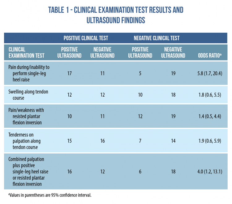
22 participants (42%) had grayscale changes in the tibialis posterior tendon on ultrasound imaging. Moderate associations were identified between ultrasound findings and two of the clinical tests – the single-leg heel raise; and combined palpation and loading test. See Table 1 for these findings.
Limitations
It would have been ideal to examine the relationship of ultrasound imaging findings to combinations of clinical tests (as would be performed in the clinic), but the sample size of the study did not provide the opportunity to explore this.
Clinical Implications
Reliability is an important consideration in order for a test to provide useful information in the clinic. The clinical tests of TPT that were evaluated in this study demonstrated moderate to substantial levels of inter-rater reliability.
The single-leg heel raise test, and combination of palpation with a loading test, were moderately associated with grayscale changes on imaging, but with low precision (indicated by the wide confidence interval of the odds ratio). This suggests that changes on ultrasound cannot be used as a surrogate for clinical tests for TPT.
This study joins existing literature to suggest that imaging findings should be considered together with clinical presentation and not in isolation. Imaging can provide information about the presence and extent of structural changes in the tendon, but this should be interpreted in the context of presenting features such as the location of pain and aggravating factors.
Here’s what this review looks like in our July issue.
Do you want to save time by not having to wade through endless piles of studies?
Let us do the hard work for you!
Every month we summarise 12 of the most recent and clinically relevant studies in physio, for instant application in the clinic.
Learn more about these Research Reviews HERE
Here are the 11 other studies we’ve reviewed in our July issue just published:
- Shoulder Strengthening for Subacromial Impingement
- Hip/Groin Muscle Strength + Symptoms in Football Players
- How Expectations Shape Pain
- Mechanical Effects of Stretching on Fascia + Muscle
- Effect of Strength Training on Pain in Knee OA
- Core-Based Exercise for Scoliosis
- Step Rate and Risk of Bone Stress Injury in Runners
- Do Injury Prevention Programs Really Work?
- Social Determinants of Health and Physiotherapy Use
- Muscle Power Predicts Mobility in Older Adults
- Pre-Season Screening of Eccentric Hamstring Strength
📚 Stay on the cutting edge of physio research!
📆 Every month our team of experts break down clinically relevant research into five-minute summaries that you can immediately apply in the clinic.
🙏🏻 Try our Research Reviews for free now for 7 days!

Don’t forget to share this blog!
Related blogs
View allElevate Your Physio Knowledge Every Month!
Get free blogs, infographics, research reviews, podcasts & more.
By entering your email, you agree to receive emails from Physio Network who will send emails according to their privacy policy.
Leave a comment
If you have a question, suggestion or a link to some related research, share below!