Plantar Plate injury, assessment and management – by Nick Knight

The following blog is by Nick Knight and was featured on Tom Goom’s website, enjoy!
Plantar plate injuries are easily missed and probably under diagnosed here in the UK. I think part of the reason for this is a lot of people don’t know what the plantar plate is. It comes back to the old saying, if you are not looking for it, you will never find it! Quite often it is grouped together with the term metatarsalgia, however this it not a diagnoses, rather just a description for pain in the ball of the foot. So what is the plantar plate? The plantar plate is a deep fibrocartilaginous structure which originates from metatarsal head and attaches to the proximal phalanx through the joint capsule within the forefoot. Its role is to help stabilize the metatarsophalangeal joints (MTPJ), along with a couple of other structures. The plantar plate also acts as an attachment site for the plantar fascia, so if you load the foot, the medial arch lengthens, the plantar fascia tightens, this engages the plantar plate to plantar flex the proximal phalanx, until the toe reaches the ground. This is a simplification of a complex process and is commonly known as the reverse windlass mechanism.1,2
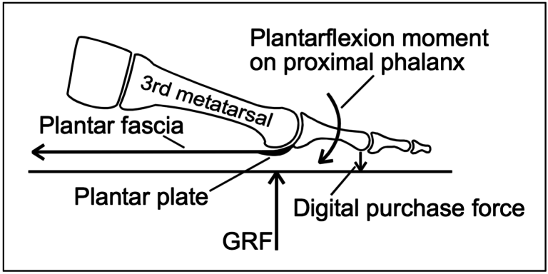
Fig 1: Permission given by K Kirby3
Stainsby4 described a simple yet effective way in testing the reverse windlass mechanism called the ‘footstool edge test’ (Fig 2) which involves standing on the edge of a stool at the MTPJ level and allowing the digits to hang over the edge. In a positive reverse windlass the proximal phalanges go into plantar flexion. If the proximal phalanges are in the same position on the ‘footstool edge test’ as to relaxed stance, this shows a non-functioning reverse windlass mechanism, which could be indicative of a plantar plate rupture.9

Figure 2: A – normal functioning reverse windlass B – non-functioning reverse windlass mechanism
So what causes a plantar plate injury and how common are they?
Plantar plate injury may be one of the most common causes of second MTPJ pain.5 The increase in the number of plantar plate tears and ruptures being diagnosed is thought to be due to advances in imaging.6,7 There are many contributing factors. The first is any activity that expose the MTPJ to repetitive and excessive dorsiflexion (resulting in increased metatarsal GRF), so think about jumping and running and, in clinic, I tend to see more of these plantar plate injuries in forefoot runners.
There are a few biomechanical causes that will increase the load through the plantar plate including hallux valgus (bunions), as the function through the 1st MTPJ is reduced, then we get what is known as low gear propulsion and increase loading through lesser MTPJs, typically the 2nd, 1st, then 3rd and so on. Also having an irregular metatarsal length, for example if you have a long 3rd metatarsal, can expose the plantar plate to increased load, as can external factors like high heels.
Basically anything that will result in excessive dorsiflexion or ground reaction forces at the MTPJs may increase plantar plate loading.
How does a plantar plate injury present?..
- The patient will complain of pain on the dorsal and plantar aspects of the MTPJ, usually described as an ache or bruising.
- Mild odeama may be present along with a episode of trauma, however trauma is not essential as plantar plate injuries are typically chronic overuse injury
- Weight bearing activities increase pain – especially dancing, forefoot running, barefoot walking etc
- Rest / non weight bearing reduces pain.
- High heels or flexible footwear increases pain
- Reduced plantar flexion strength – The ‘Digital Purchase’ test
- Pain, oedema and positive Digital Lachmans (Anterior Draw) / Vertical Stress.
- Quick way to do this, put a piece of paper under the apex of the effected toe and ask the patient to try and stop you pulling the paper away, in a plantar plate injury you will notice the paper is pulled away much more easily.
- Floating toe, if late stage hammer toe, or Churchill sign may be present (Figure 3 and 4)
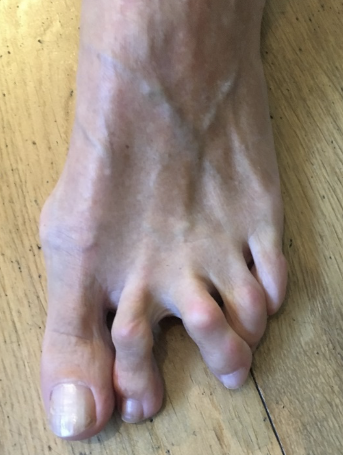
Figure 3: Churchill Sign
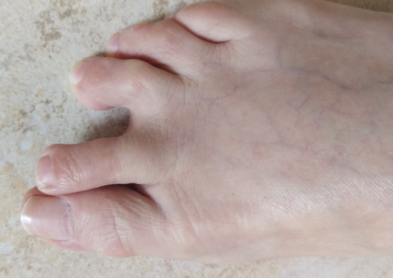
Figure 4: Churchill Sign
Digital Lachmans / Vertical Stress Test (Fig 5)
Same style of test to assess ACL tears, helps to assess integrity of the plantar pate, it is quick, easy and a simple test to perform. Stabilise the head of the metatarsal with one hand, using the other hand stabilise the base of the proximal phalanx, apply a vertical force, we are looking for pain and any translocation, it is important to remember this is different from dorsiflexion of the digit.
One study showed sensitivity of 80.6% (positive vertical stress test with tear identified intraoperatively) and a specificity of 99.8% (negative vertical stress test with intact plantar plate intraoperatively)8
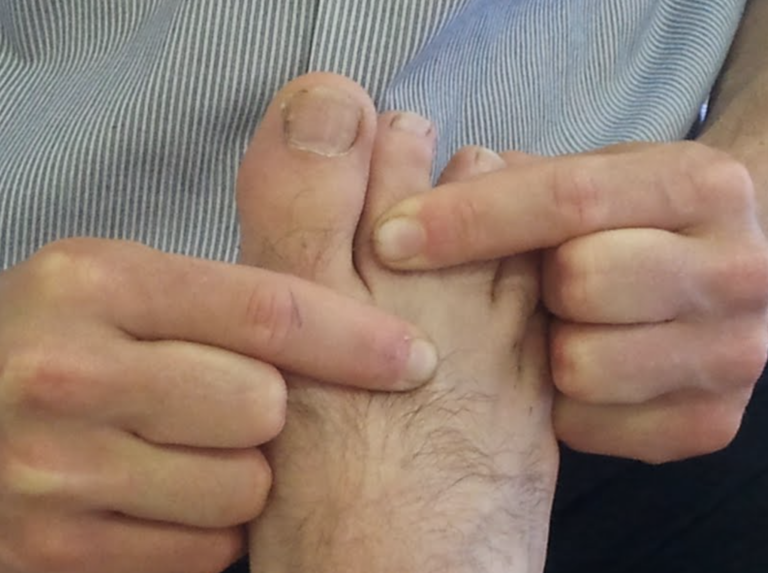
Figure 5: A Stage 0 Vertical stress test.
There are 2 scoring systems one by Thompson and Hamilton9 and the other Yu and Judge10
Thompson and Hamilton9
- Stage 0, there is no dorsal translocation present of the proximal phalanx.
- Stage 1 the base of the phalanx, will not dislocate, however may sublux
- Stage 2 the base of the phalanx can be dislocated.
- Stage 3 the phalanx base is in a fixed dislocated position
Yu and Judge10
- Stage 1 mild odema on the plantar MTPJ with dorsal odema often present as well. Tenderness is present on palpation, however no anatomical malalignment.
- Stage 2 moderate odema is present with noticeable deviation.
- Stage 3 odema present around the entire MTPJ with deviation and possible dislocation/subluxation, the odema will reduce however the deformities will remain.
I think the best way to describe the 2 different methods of testing, would be that the Thompson and Hamilton test best describes the integrity of the plantar plate at any given time, whereas the Yu and Judge test describes different stages based on clinical findings on the time of examination.
The role of Imaging
There is still some debate as to whether an MRI scan or ultrasound scan is best for detecting plantar plate injuries. As we know ultrasound is cheaper, however it is user dependent, whereas MRI scan is more expensive but we can also get an overall picture of the structures within that area as well. A recent systematic review showed a sensitivity of 95% in MRI scans and 93% in ultrasound scan, whilst specificity was 54% in MRI and 33% in ultrasound11
X-ray in weight bearing (lateral or oblique views) will show subluxation dorsally of the proximal phalanx on the metatarsal head, an anterior posterior view will show a transverse deformity as well. An x-ray will also rule out other bony pathologies.12
Interestingly one study did examine 160 asymptomatic and 160 symptomatic plantar plates, they did find that 35% of the asymptomatic group had a plantar plate injury.13 This then raises the question then shall we treat the asymptomatic tears? We can approach of 2 ways, one that it is currently asymptomatic leave it alone and treat the patient not the scan, or secondly, we offer conservative measures to start with, as we know that if the plantar plate ruptures or becomes dysfunctional, floating toes, and hammer toes deformities may start to appear which may become more difficult to manage later.
Treatment
The aim of treatments, like most musculoskeletal pathologies is about managing the load. Essentially we want to try and reduce the ground reaction forces under the affected metatarsal head and reduce the plantar flexion moment of the metatarsal and the dorsiflexion of the phalanx.
The treatment can include
- No barefoot walking / activity modification
- Footwear advice / Air cast boot – we want to look at using a stiff soled shoe, or reducing the heel height of a shoe, so footwear like high heels and the flexible minimalist type shoes tend to aggravate a plantar plate injury, the same goes for open toe shoes and flip-flops, as you must claw your toes to keep these on which again increases the ground reaction force underneath the metatarsal.
- Stretching / Strengthening – thinking about the mechanics of the foot, if there is tightness within the calf muscles, in turn could result in early and increased loading through the forefoot, and if you are unable to get adequate dorsiflexion due to calf tightness, then the foot may pronate to compensate for this, which in turn could increase the loading through the lesser MTPJ’s. It is important also to work on strengthening the muscles within the foot.
- Strapping, can be very helpful in reducing pain, using a rigid zinc oxide tape and pulling the toe into a plantar flexed position to help offload a plantar plate (Fig 6).
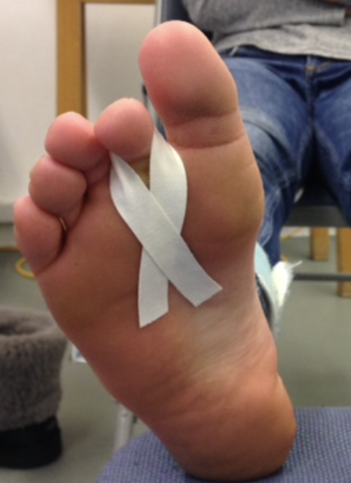
Figure 6: Strapping for plantar plate injury
Orthoses, can be a useful way to help offload the affected plantar plate, using a metatarsal dome just proximal to the affected metatarsal head and then a sulci extension with a cut out around the MTPJ region (Fig 7). Combining the orthoses, taping and footwear advice can be quite an effective way of offloading the affected plantar plate, whilst the patient reduces sporting activities.
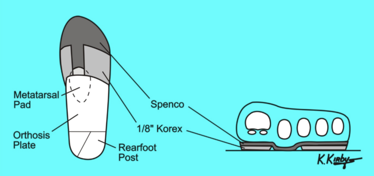
Figure 7: Permission given by K Kirby3
Steroid injection – A staggered course of steroid injections reaching a maximum of once ever 1 to 3 months and a maximum 3 in a 12 month period has been shown to effective and safe, however repeated intra-articular injections has been shown to result in dislocation of the MTPJ.12,14,15 it has also been suggested that injections into a ligament resulted in destruction of fibrocytes and reduction in tensile strength for up to 1 year16 which in turn may result in further damage a possible rupture.
A recent case study showing a patient with a plantar plate tear, was managed using conservative measures, consisting of taping, activity modification and the use of a Darco boot over a 6 month period, and progressing to stiffed shoe and orthoses (however the orthoses where not described) and stopped taping. At the 1 year mark, the patient was pain free with no toe deformity, and on MRI the plantar plate has healed.
So what’s my treatment plan?
- No bare foot walking for 6 weeks (minimum)
- To wear stiff soled shoes
- Strapping of digit changing every 72 hours
- Activity modification
- Orthoses as described as above, plus any other modifications required
- Stretching and Strength work – Distal and proximal
Review regularly and, if conservative measures fail, then it requires a referral to my surgical colleagues. Hopefully when you next see a hammer toe you will realise that is quite a complex process! The recent case study published, provides a nice base of more research to be conducted, looking at if we can prevent planter plates rupturing and allow to heal, in turn reducing likelihood of lesser toes deformities.
Check out Nick’s website and be sure to follow him on Twitter via @NKSportsPod.
Want to master knee OA?
Dr Allison Ezzat has done a Masterclass lecture series for us!
“Knee Osteoarthritis Essentials: Practical Strategies for Clinicians”
You can try Masterclass for FREE now with our 7-day trial!

Don’t forget to share this blog!
Related blogs
View allElevate Your Physio Knowledge Every Month!
Get free blogs, infographics, research reviews, podcasts & more.
By entering your email, you agree to receive emails from Physio Network who will send emails according to their privacy policy.
Leave a comment
If you have a question, suggestion or a link to some related research, share below!